
The Brain – Facts, Parts and Layers
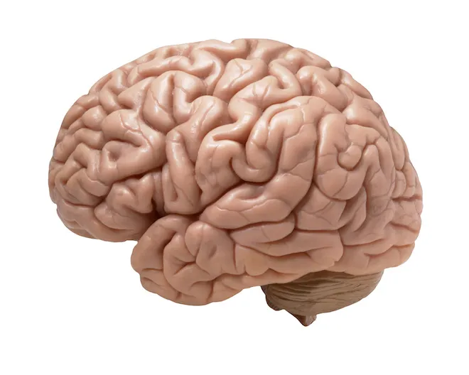
Human Brain
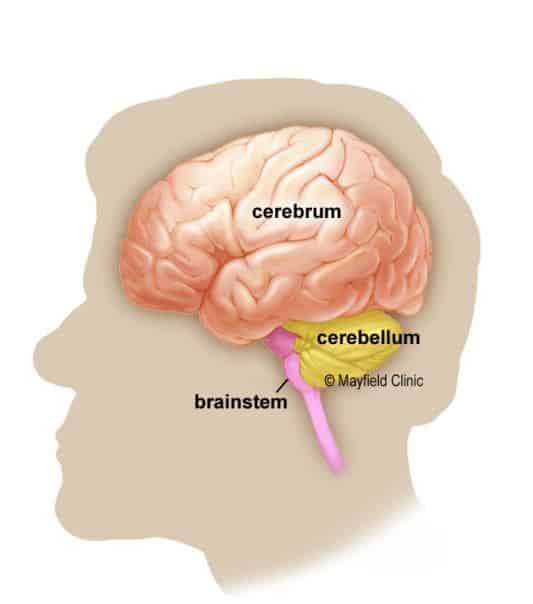
The human brain is an amazing three-pound organ that controls all functions of the body, interprets information from the outside world, and embodies the essence of the mind and soul. Intelligence, creativity, emotion and memory are a few of the many things governed by the brain. Protected within skull, the brain is composed of the cerebrum, cerebellum and brainstem.
The brain receives information through our five senses : sight, smell, touch, taste and hearing – often many at one time. It assembles the messages in a way that has meaning for us, and can store that information in our memory. The brain controls our thoughts, memory and speech, movement of the arms and legs, and the function of many organs within our body.
The central nervous system (CNS) is composed of the brain and spinal cord. The peripheral nervous system (PNS) is composed of spinal nerves that branch from the spinal cord and cranial nerves that branch from the brain.
Brain Facts
– Composition of the brain: 78% Water, 12% Lipids, 8% Proteins, 1% Carbs, 2% Soluble organics and 1% Salt.
– The slowest speed at which information travels between neurons is 260 mph (416 km/h).
– The brain can stay alive for 4-6 mins without oxygen; after that, cells begin to die.
– More electrical impulses are generated in one day by a single human brain than by all the telephones in the world.
– Around 70,000 thoughts are produced by a human brain in any single day.
– 89.04% is the percentage of people who report normally writing with their right hand, 10.6% with their left, and 0.34% with either hand.
– The brain is composed majorly of three parts, cerebrum, cerebellum and brainstem.
Difference between Male and Female Brain
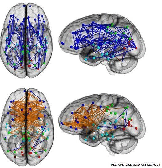
- Male brains see most of their neuron activity firing front-to-back within each hemisphere of the brain, with little overlap. Girls’ brains have less hemisphere isolation and instead have much more cross-hemisphere neuron activity. This explains why girls can multitask better.
- Men’s brain are, on average, 10% larger than women’s, and consequentially weigh slightly more, 3 lbs to 2 3/4 lbs. These size differences have been found repeatedly, but they emerge only when comparing large numbers of people, so some women’s brains are larger than the average whereas some men’s are smaller. These differences partly reflect the fact that men are generally bigger and taller than women, but they are not in any way related to differences in intelligence.
- Men and women’s brains also differ in overall composition. Male brains contain about 6.5 times more grey matter than women, while female brains have more than 9.5 times as much white matter. The frontal and the temporal areas of the brain cortex are larger in volume in women.
- The Amygdala is larger in males; the Hippocampus is activated on its right side in males yet on its left side in females.
- Stress has been found to induce an increase in serotonin and dopamine levels within the amygdala in males, but not in females. In females, both short-term and long-stress have been found to actually enhance spatial memory while under duress.
Why did the brain need sleep ?
- To process the day’s events and exposure to knowledge. When we sleep, the brain continues to process the input from the day and make sense of it. For instance, in a math concept you learned Monday was difficult and you see uncertain, brain scans have revealed that while you sleep the area of the brain involved with math is active, and in the morning what was difficult on Monday seems easier to understand on Tuesday.
- When you sleep, the upper spinal cord releases a fluid which serves to “flush out” toxic waste products which cells produce with daily use; effectively cleaning our the brain. It removes inflammatory, toxins and protein plaque buildup (associated with Alzheimer’s); the waste is flushed out into the bloodstream, through which it is eventually carried to the liver for detoxification.
- Discredited Theory : It used to be thought that sleep helps animals conserve energy by forcing a period of rest. But this theory is deemed unlikely since the sleeping brain uses up almost as much energy as awake brain.
Parts of Brain
Cerebrum
It is the largest part of the brain (83% of brain mass) and is composed of right and left hemispheres. It performs higher functions like interpreting touch, vision and hearing, as well as speech, reasoning, emotions, learning, and fine control of movement.
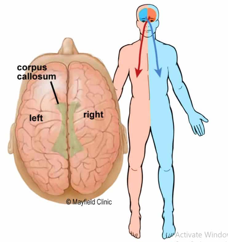
Right Brain – Left Brain
The cerebrum is divided into two halves : the right and left hemispheres. They are joined by a bundle of fibres called the corpus callosum that transmits messages from one side to the other.
Each hemisphere controls the opposite side of the body. If a stroke occurs on the right side of the brain, your left arm or leg may be weak or paralysed. Not all functions of the hemispheres are shared.
(i) Left Brain
In general, the left hemisphere controls speech, comprehension, arithmetic, and writing. The left hemisphere is dominant in hand use and language in about 92% of people. It helps children acquire language, make the connections between the sounds heard and things seen or otherwise experienced, and seems to control speech in 96% of children. The left brain is good at exact and precise thought processes, and the pre-motor areas of this region deals with grammar. The left hemisphere of the brain has 186 milllion more neurons than the right hemisphere.
(ii) Right Brain
The right hemisphere controls creativity, spatial ability, artistic, and musical skills. The right side is involved in interpreting and generating speech with meanings. The right brain is apt at making broad and sweeping understandings and it can also decipher meaning by ‘the way you said it’, the point of emphasis and comprehends humor. People with Dyslexia have a slightly larger Right Hemisphere.
Cerebellum
It is located under the cerebrum (11% of brain mass). It is also called “Little Brain”. Its function is to coordinate muscle movements, maintain posture and balance.
Brainstem
It acts as a relay center connecting the cerebrum and cerebellum to the spinal cord. It is located between diencephalon and the spinal cord. It is composed of three parts: Mid Brain, Pons and Medulla Oblongata. It performs many automatic functions such as breathing, heart rate, body temperature, wake and sleep cycles, digestion, sneezing, coughing, vomiting, and swallowing.
(a) Pons
The pons serves as a bridge to connect areas of the brain to one another. The pons contains areas that relay signals for voluntary movements, equilibrium information from the inner ear, and areas that (together with the medulla oblongata) help control breathing. This maintains a role in sleep, particularly in terms of being conscious (awake) or not, regulate consciousness and is associated with the sense of higher pupose.
“Locked-in Syndrome”, a condition in which a patient is aware and awake but cannot move or communicate due to complete paralyses except for the eyes, occurs when a lesion damages the Pons.
Gray Matter and White Matter
The neuron highways! Using the concept of a computer as an analogy: gray matter can be thought of as the actual computers themselves, whereas the white matter represents the network cables connecting the computers together.
The brain can adapt to white matter damage. Unlike gray matter, which peaks in development in a person’s twenties, the white matter continues to develop and peaks about middle age.
In terms of oxygen consumed, 6% will be used by the brains white matter and 94% by the gray matter. The development of Multiple Sclerosis as well as protein build up associated with Alzheimer’s both affect white matter.
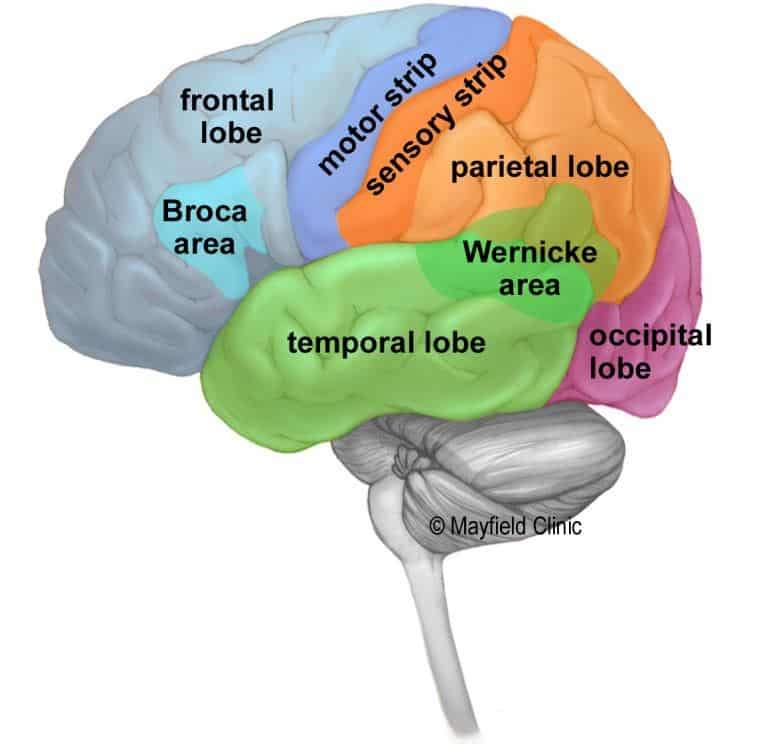
Lobes of the Brain
The cerebral hemispheres have distinct fissures, which divide the brain into lobes. Each hemisphere has 4 lobes: Frontal, Temporal, Parietal, and Occipital. Each lobe may be divided, once again, into areas that serve very specific functions. It is important to understand that each lobe of the brain does not function alone. These are very complex relationships between the lobes of the brain and between the right and left hemispheres.
Language
In general, the left hemisphere of the brain is responsible for language and speech and is called the ‘dominant’ hemisphere. The right hemisphere plays a large part in interpreting visual information and spatial processing. In about one third of people who are left handed, speech function may be located on the right side of the brain. Left-handed people may need special testing to determine if their speech centre is on the left or right side prior to any surgery in that area.
Cortex
The surface of the cerebrum is called the cortex. It has a folded appearance with hills and valleys. The cortex contain 16 billion neurons (the cerebellum has 70 billion = 86 billion total) that are arranged in specific layers. The nerve cell bodies color the cortex grey-brown giving it its name – gray matter. Beneath the cortex are long nerve fibers (axons) that connect brain areas to each other – called white matter.
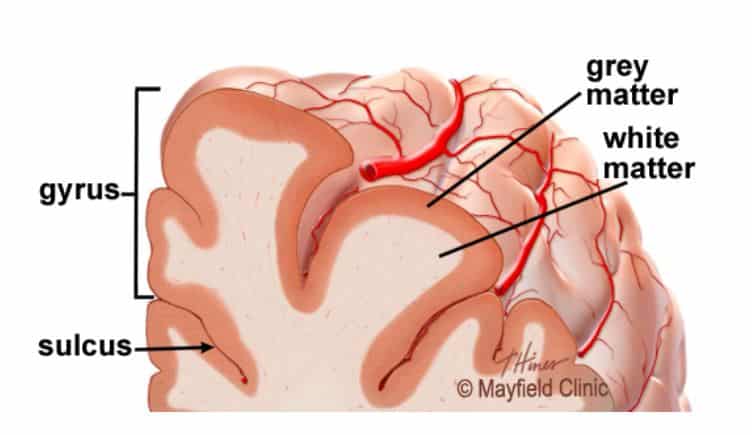
The folding of the cortex increases the brain’s surface area (approx three times) allowing more neurons to fit inside the skull and enabling higher functions. Each fold is called a gyrus (2-4 mm thick), and each groove between folds is called a sulcus. There are names for the folds and grooves that help define specific brain regions.
Deep Structures
Pathways called white matter tracts connect areas of the cortex to each other. Messages can travel from one gyrus to another, from one lobe to another, from one side of the brain to the other, and to structures deep in the brain.
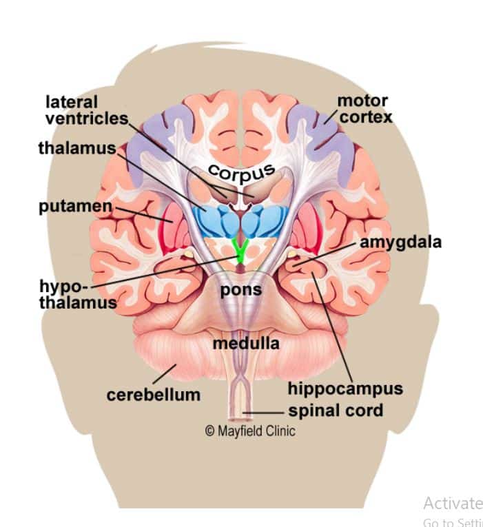
Hypothalamus
It is located in the floor of the third ventricle and is the master control of the autonomic system. It plays a role in controlling behaviours such as hunger, thirst, sleep, sexual response, as well as circadian (24-hour cycle) rhythms. It also regulates body temperature, blood pressure, emotions, and secretion of hormones. Failure to get 8-9 hours of sleep each night can disrupt teens circadian rhythm ‘clock’.
Medulla Oblongata
This governs involuntry processes, such as breathing, swallowing, defecation (i.e, need to use toilet), digestion and heart rate. It is located on the lower half of the brain stem, next to the pituitary gland.
Pituitary Gland
It lies in a small pocket of bone at the skull base called the sella turcica and is of size of a pea. The pituitary gland is connected to the hypothalamus of the brain by the pituitary stalk. Known as the “master gland”, it controls other endocrine glands in the body. It secretes hormones that control sexual development, promote bone and muscle growth, and respond to stress.
Pineal Gland
It is located behind the third ventricle. It helps regulate the body’s internal clock and circadian rhythms by secreting melatonin. It has some role in sexual development.
Thalamus
It serves as a relay station for almost all information that comes and goes to the cortex. It plays a role in pain sensation, attention, alertness and memory. It also is suspected to play a function in muscle control.
Basal Ganglia
It includes the caudate, putamen and globus pallidus. These nuclei work with the cerebellum to coordinate fine motions, such as fingertip movements.
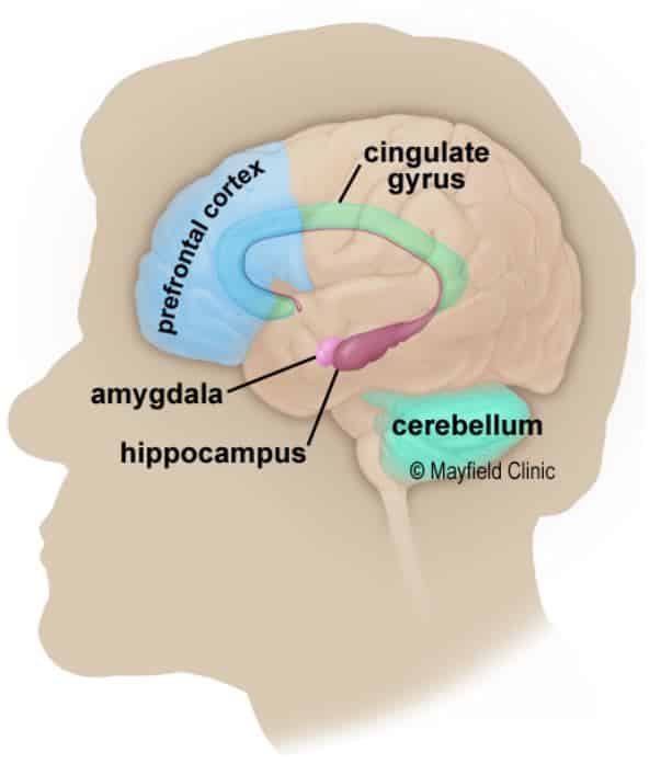
Limbic System
It is the center of our emotions, learning, and memory. Included in this system are the cingulate gyri, hypothalamus, amygdala (emotional reactions) and hippocampus (memory).
Amygdala
It helps in storing and classifying emotionally-charged memories. It plays a significant role in producing emotions, especially fear and jealousy. In teenagers, the Amygdala is more active than the Frontal Lobe; individuals with an enlarged Amygdala have been diagnosed with manic-depressive disorder.
Memory
Memory is a complex process that includes three phases: encoding (deciding what information is important), storing, and recalling. Different areas of the brain are involved in different types of memory. Your brain has to pay attention and rehearse in order for an event to move from short-term to long-term memory – called encoding.
Hippocampus
Its primary role is memory formation, classifying information and long-term memory. Leading thought hypothesizes that this area also intuitively informs a person of their place within an environment.
People with extensive damage to this part of the brain may suffer amnesia. Furthermore, in Alzheimer’s Disease, this area is among the first to suffer damage.
Short-term Memory
It is also called working memory, occurs in the prefrontal cortex. It stores information for about one minute and its capacity is limited to about 7 items. For example, it enables you to dial a phone number someone just told you. It also intervenes during reading, to memorize the sentence you have just read, so that the next one makes sense.
Long-term memory
It is processed in the hippocampus of the temporal lobe and is activated when you want to memorise something for a longer time. This memory has unlimited content and duration capacity. It contains personal memories as well as facts and figures.
Skill Memory
It is processed in the cerebellum, which relays information to the basal ganglia. It stores procedural learned memories like tying a shoe lace, playing an instrument, or riding a bike.
Ventricles and Cerebrospinal fluid
The brain has hollow fluid-filled cavaties called ventricles. Inside the ventricles is a ribbon-like structure called the choroid plexus that makes clear colorless cerebrospinal fluid (CSF). CSF flows within and around the brain and spinal cord to help cushion it from injury. This circulating fluid is constantly being abrorbed and replenished.
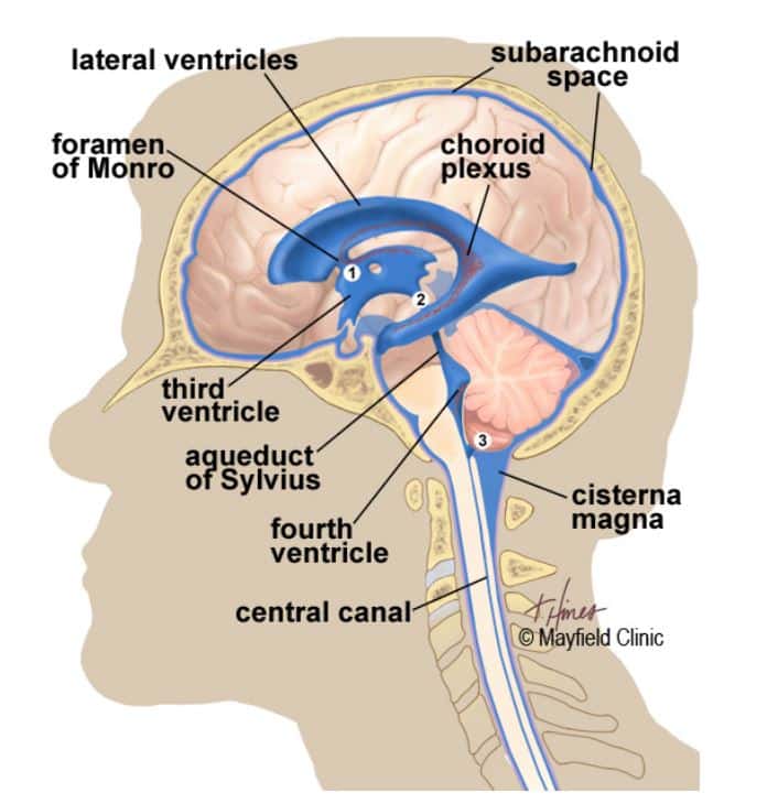
There are two ventricles deep within the cerebral hemispheres called the lateral ventricles. They both connect with the third ventricle through a separate opening called the foramen of Monro. The third ventricle connects with the fourth ventricle through a long narrow tube called the aqueduct of Sylvius. From the fourth ventricle, CSF flows into the subarachnoid space where it bathes and cushions the brain. CSF is recycled (or absorbed) by special structures in the superior sagittal sinus called arachnoid villi.
A balance is maintained between the amount of CSF that is absorbed and the amount that is produced. A disruption or blockage in the system can cause a build up of CSF, which can cause enlargement of the ventricles (hydrocephalus) or cause a collection of fluid in the spinal cord (syringomyelia).
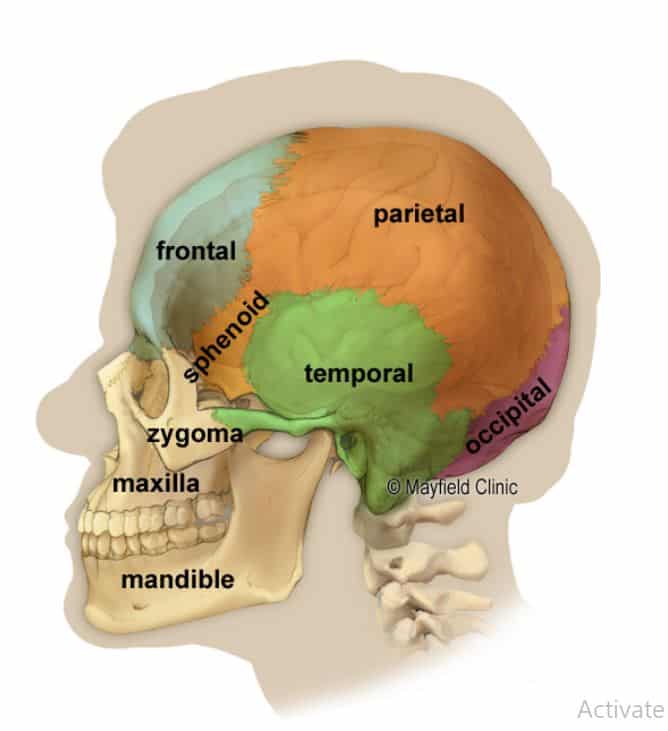
Skull
The purpose of the bony skull is to protect the brain from injury. The skull is formed from 8 bones that fuse together along suture lines. These bones include the frontal, parietal (2), temporal (2), sphenoid, occipital and ethmoid. The face is formed from 14 paired bones: the maxilla, zygoma, nasal, palatine, lacrimal, inferior nasal conchae, mandible, and vomer.
Inside the skull are three distinct areas: anterior fossa, middle fossa, and posterior fossa. Doctor sometimes refer to a tumor’s location by there terms e.g. middle fossa meningioma,
Similar to cables coming out the back of a computer, all the arteries, veins and nerves exit the base of the skull through holes, called foramina. The big hole in the middle (foramen magnum) is where the spinal cord exits.
Cranial Nerves
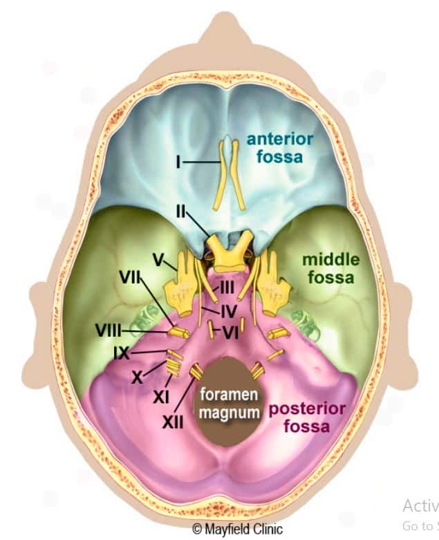
The brain communicates with the body through the spinal cord and twelve pairs of cranial nerves. Ten of the twelve pairs of cranial nerves that control hearing, eye movement, facial sensations, taste, swallowing and movement of the face, neck, shoulder and tongue muscles originate in the brainstem. The cranial nerves for smell and vision originate in the cerebrum.
Name and main function of the twelve cranial nerves are :-
- Olfactory Smell (I)
- Optic Sight (II)
- Oculomotor Moves Eye, Pupil (III)
- Trochlear Moves Eye (IV)
- Trigeminal Face sensation (V)
- Abducens Moves eye (VI)
- Facial Moves face, Salivate (VII)
- Vestibulocochlear Hearing and Balance (VIII)
- Glossopharyngeal Taste, Swallow (IX)
- Vagus Heart rate, Digestion (X)
- Accessory Moves head (XI)
- Hypoglossal Moves tongue (XII)
Layers of the Brain
The brain is surrounded by three layers of connective tissue membrane called meninges. These layers cover and protect the brain and blood vessels, contain cerebrospinal fluid, and create partitions in the skull. From the outermost layer inward they are: the dura mater, arachnoid mater, and pia mater.
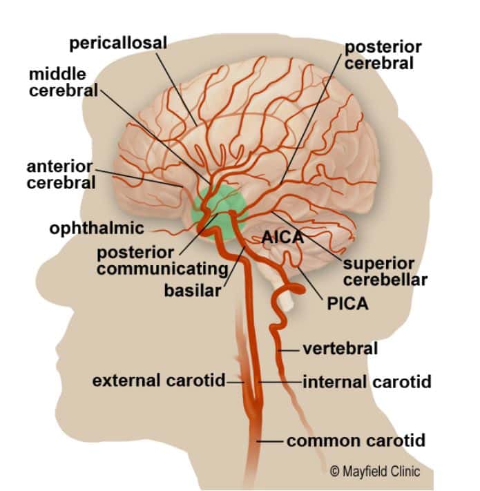
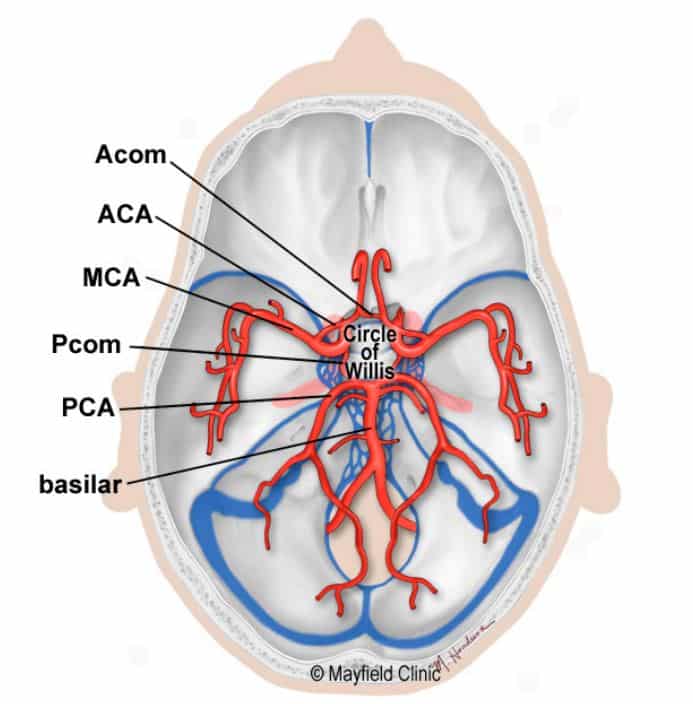
Blood Supply to the Brain
To function correctly, our brains require a continuous supply of well-oxygenated blood. After just 10 seconds without it, we lose consciousness. After a few minutes, irreversible damage usually begins to manifest. To account for this dependency, neural tissues are supplied by a dense meshwork of blood vessels.
Blood is carried to the brain by two paired arteries, the carotid arteries known as anterior circulation and the vertebral arteries known as posterior circulation.
Cells of the Brain
The brain is made up of two types of cells: Nerve cells (neurons) and glia cells.
Nerve Cells
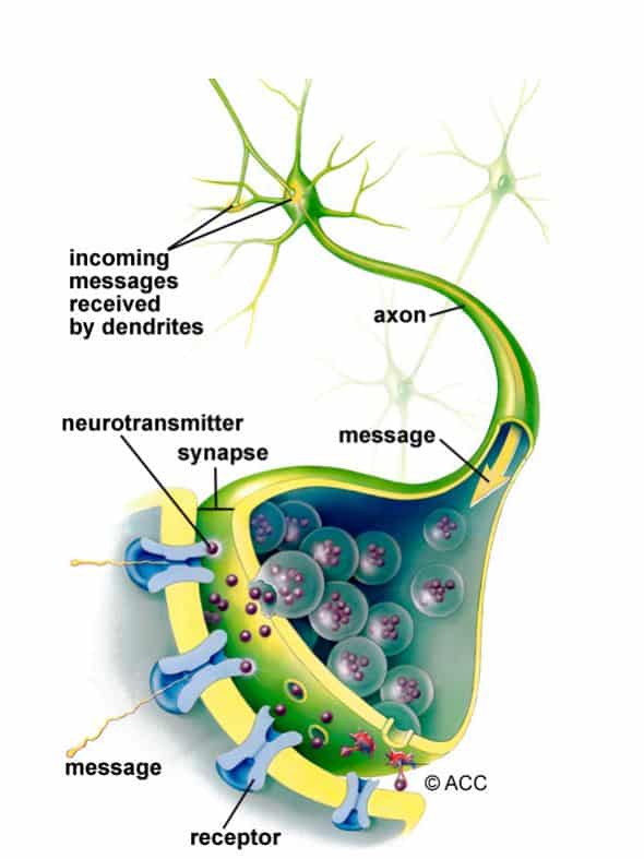
There are many sizes and shapes of neurons, but all consist of a cell body, dendrites and an axon. The neuron conveys information through electrical and chemical signals. Try to picture electrical wiring in your home. An electrical circuit is made up of numerous wires connected in such a way that when a light switch is turned on, a light bulb will beam. A neuron that is excited will transmit its energy to neurons within its vicinity.
Neurons transmit their energy, or “talk”, to each other across a tiny gap called a synapse. A neuron has many arms called dendrites, which act like antennae picking up messages from other nerve cells. These messages are passed to the cell body, which determines if the message should be passed along. Important messages are passed to the end of the axon where sacs containing neurotransmitters open into the synapse. The neurotransmitter molecules cross the synapse and fit into special receptors on the receiving nerve cell, which stimulates that cell to pass on the message.
Glia Cells
Glia (Greek word meaning Glue) are the cells of the brain that provide neurons with nourishment, protection, and structural support. There are about 10 to 50 times more glia than nerve cells and are the most common type of cells involved in brain tumors.
– Astroglia or astrocytes are the caretakers – they regulate the blood brain barriers, allowing nutrients and molecules to interact with neurons. They control homeostasis, neuronal defense and repair, scar formation, and also affect electrical impulses.
– Oligodendroglia cells create a fatty substance called myelin that insulates axons – allowing electrical messages to travel faster.
– Ependymal cells line the ventricles and secrete cerebrospinal fluid (CSF).
– Microglia are the brain’s immune cells, protecting it from invaders and cleaning up debris. They also prune synapses.